RadAnat 2026: Online Workshop on Radiological Anatomy
Precision Anatomy for Modern Diagnostic Imaging
20 April – 30 April 2026 | 7:00 PM IST | Online | Certificate
India – 7:00 PM IST | Saudi Arabia – 4:30 PM | Dubai – 5:30 PM | Singapore – 10:30 PM | Australia – 11:30 PM | New York – 9:30 AM
About the Workshop
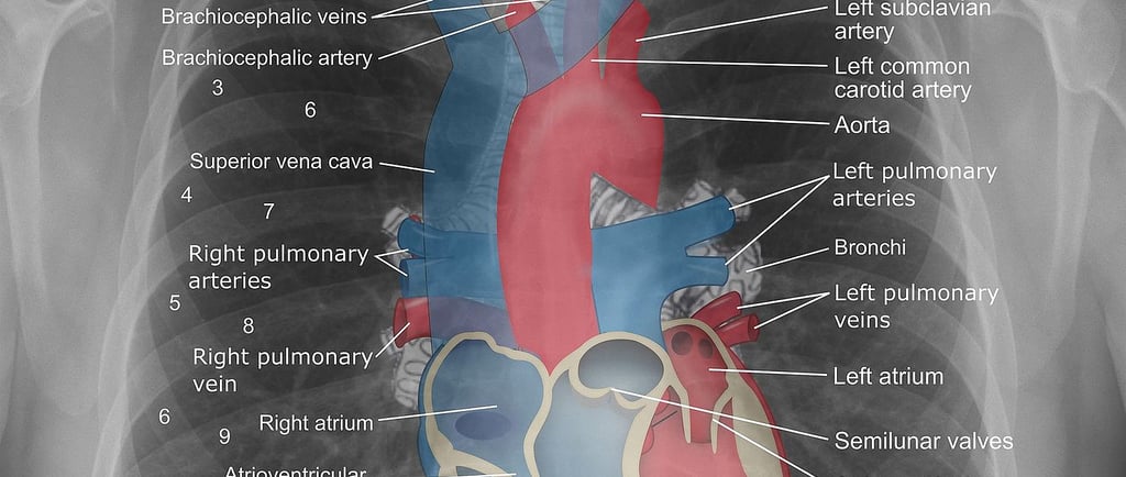
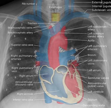
This online certificate workshop RadAnat 2026: Online Workshop on Radiological Anatomy is designed to help participants understand how human anatomy is visualized through modern medical imaging techniques. The program bridges the gap between textbook anatomy and real-world diagnostic imaging using X-ray, CT, MRI, and ultrasound. Through expert-led interactive sessions, participants will learn to identify anatomical structures, interpret radiological images, and understand positioning principles used in clinical imaging. The workshop emphasizes image-based learning, real case examples, and practical understanding essential for radiology and allied healthcare fields.
What You Will Learn
Fundamentals of radiological anatomy and its clinical applications
Differences between gross anatomy and imaging anatomy
Understanding tissue densities and image contrast
Anatomical planes and radiological terminology
Identification of anatomical landmarks on X-ray, CT, and MRI
Correlation between surface anatomy and radiographic anatomy
Standard radiographic views (PA, AP, lateral, oblique)
Patient positioning techniques in diagnostic imaging
Skeletal anatomy (axial and appendicular) using radiological images
Recognition of normal vs pathological structures
Course Features
Live online interactive sessions
e-Certificate upon completion
Access to session recordings
Lecture PPTs and study materials
Software-based and image-based learning
Real radiograph and CT case discussions
Expert faculty from Radiology & Anatomy
Learning Outcomes
Understand the role of radiological anatomy in clinical diagnosis
Identify anatomical landmarks on X-ray, CT, and MRI images
Interpret normal radiographic appearances of body structures
Apply anatomical knowledge in diagnostic imaging
Understand patient positioning for accurate imaging
Recognize skeletal structures and anatomical variations
Who Can Join
B.Sc. / M.Sc. Radiology & Medical Imaging students
Allied Health Science & Paramedical students
Radiology interns and imaging technicians
Faculty in medical and allied sciences
Beginners interested in radiological anatomy
Anyone aiming to build a career in diagnostic imaging
Course Fee
Students (India): ₹1499 INR
Faculty / Professionals: ₹1999 INR
International Participants: $85 USD
Workshop Module
WEEK 1: Fundamentals of Radiological Anatomy
Introduction to Radiological Anatomy
Scope and applications in diagnostic imaging
Comparison between gross anatomy and radiological anatomy
Understanding tissue densities and image contrast
Anatomical and Radiological Terminology
Body planes (sagittal, coronal, transverse)
Anatomical positions and movement terminology
Radiographic symbols, abbreviations, and orientation markers
Identification of Key Anatomical Landmarks
X-ray, CT, and MRI interpretation basics
Correlation of surface anatomy with imaging anatomy
Normal radiographic appearances (head, chest, abdomen)
Standard Radiographic Positions and Projections
PA, AP, lateral, and oblique views
Patient positioning techniques
Learning Outcomes (Week 1)
Explain the importance of radiological anatomy
Identify body planes and anatomical landmarks
Understand imaging orientation and positioning
Assessment (Week 1)
Quiz: Body Planes & Positions
Radiograph Spot Test: Landmark Identification
WEEK 2: Skeletal Radiological Anatomy (Axial & Appendicular Skeleton)
A. Axial Skeleton
Skull: cranial & facial bones, sinuses, sutures
Vertebral column: cervical, thoracic, lumbar, sacral regions
Thoracic cage: ribs, sternum, costal cartilage
B. Appendicular Skeleton
Upper limb: shoulder girdle, arm, forearm, wrist, hand
Lower limb: pelvic girdle, thigh, leg, ankle, foot
Identification of joints, alignment, and ossification centers
Learning Outcomes (Week 2)
Identify bones and joints on radiographs and CT images
Recognize skeletal landmarks and anatomical structures
Differentiate normal anatomy from pathological findings
Assessment (Week 2)
X-ray Labeling Activity
CT Bone Anatomy Quiz